XDH Gene Mutation is the Underlying Cause of Classical Xanthinuria: A Second Report

Xanthinuria is an autosomal recessive disorder that presents itself through excessive excretion of xanthine in urine. Type I xanthinuria results from a deficiency of xanthine dehydrogenase (xdh); the xdh gene is located at chromosome 2p22-23. Type II xanthinuria results from deficiency of both XDH and aldehyde oxidase. The primary purpose of the study was to understand the molecular basis of xanthinuria through an analysis xdh mutations; as such they are dealing with type I xanthinuria.  Subtyping xanthinuria was attempted by homozygosity mapping. Mutation detection was accomplished by PCR-SSCP screening of the entire xdh gene; all 36 exons and exon-intron junctions of the patient’s gene. The gene sequence was then confirmed. The researchers determined that the xanthinuria in the patient is linked to the xdh gene mutation; as suggested by their data. As a result a new 1658insC mutation in exon 16 of the xdh gene was identified; through the analysis of the patient’s xdh gene. The researchers demonstrated the linkage of xanthinuria to the xdh locus by homozygosity mapping. Additionally, the newly identified 1658insC mutation predicted an inactivated xdh protein. They conclude by saying that their results “reinforce the notion that mutations in the xdh gene are the underlying cause of classical xanthinuria type I.”

References:

David Levartovsky, Ayala Lagziel, Oded Sperling, Uri Liberman, Michael Yaron, Tatsuo Hosoya, Kimiyoshi Ichida and Hava Peretz. Tel Aviv Sourasky Medical Center and Rabin Medical Center, Tel Aviv University, Tel Aviv, Israel; and The Jikey University School of Medicine, Tokyo, Japan. Published: 7 January 2000.

Additionally a Link to the Full Text is Provided Below:

http://www.nature.com/ki/journal/v57/n6/full/4491562a.html

Cross-Competition in Transgenic Chloroplasts Expressing Single Editing Sites Reveals Shared cis Elements

Research performed by: Anne-Laure Chateigner-Boutin and Maureen R. Hanson
While some RNA editing sites require a lot of surrounding sequences, others require a minimal amount. For each one of these individual editing sites, there are various specific trans-acting elements that are used during regulation of a different target gene. Researchers gathered that if each editing site were independent, more than 400 proteins would be necessary for successful recognition of the editing site by the organelle. In order to test this theory of independence of the editing sites in chloroplasts, the researches conducted an experiment in which they carefully observed the degree of editing in thirty-one known tobacco chloroplast editing sites in two different tobacco lines, both of which contained plastids that were homogeneously transgenic for some foreign gene. Researchers were not proven right, however, and they found that the more each gene was expressed, the less editing was occurring at other sites. When comparing the sequences around the introduced sites with sequences around the sites where editing had been altered, the researchers were able to examine discrete transgenic elements that were 5’ of the C target of editing. After seeing this result in the chloroplasts, the researchers suggested that perhaps the same sort of apparatus occurs in the mitochondria, where the same sort of known cis transgenic elements are also able to be observed 5’ of the C target of editing. After conducting an experiment to test this theory, the researchers found that transgenic elements similar to those found on one of the specific sites in the chloroplasts were also found on many mitochondrial genes. Unless there was some feedback mechanism that an increased spread of the factor to the chloroplast sites, it is not likely that the abundance of elements in the chloroplasts affected the editing in the mitochondria.
SOURCE: http://mcb.asm.org/content/22/24/8448.full.pdf+html

Primer development for the plastid region ycf1 in Annonaceae and other magnoliids

This article identifies the usability of ycf1 using primers to identify the plasid gene within the magnoliid taxa. This hypothetical chloroplast open reading frame 1, more commonly identified as ycf1, codes a region of 5,500 base pairs, and is virtually present in many plastid genomes. It is typically used  in analyzing orchids and Pinus, making the use of it in magnoliids unique and somewhat rarely studied. Using an array of species across many families, the samples were able to specifically identify the usage of ycf1. An array of primers were used to amplify various portions, to amplify various nucleotide regions. By sequencing roughly 2000 base pairs within ycf1 in over 20 individuals, the researchers were able to examine the phylogenetic utility relative to other plastid regions. The genera Deeringothamnusand Asimina (Annonaceae) was used due to its close proximity. Through various PCR deviations using different primers revealed the discrepancy of ycf1 among species, discovering the higher number of informative characters. Finally, the resulting factors proved that ycf1 proves to be easily amplified and sequenced vastly more than other common markers such as matK and trnL-F. The study indicates that ycf1 has great potential utility.


References:

Neubig, K., & Abbott, J. (2010). Primer development for the plastid region ycf1 in annonaceae and other magnoliids. American Journal of Botany, Retrieved from http://www.amjbot.org/content/97/6/e52.full

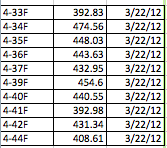
DNAs 4-33 to 4-44 Amplified atpF

Emmanuel and Archana were able to successfully detect regions of DNA from samples numbered 4-33 to 4-44 that were amplified on March 22nd. According to the AASU Genetics Blog, all of the samples (excluding one) yielded a bright band, indicating the presence of a successful amplification of that region. The only sample that was not used was 4-43. After being treated with Exonuclease, the samples that were used were incubated. The Nano-drop spectrophotometer, which measures the quantity of DNA present, was used to measure and quantify our samples so that an accurate result showing the amount of DNA present could be obtained. The whole procedure can be found at https://armstronggenetics3.wordpress.com/category/techniques/. The results, as shown in the data table below, there was a consistently high yield of DNA in all of the samples that were quantified. The first column is the sample number, the second is the quantified number (ng/ul), and the third is the data quantification was performed. The range was 392.83 ng/ul to 474.56 ng/ul, which doesn’t show that much variation in data. As always, there are possible sources of error, such as pipetting, denaturation of DNA, initial mix-up of samples, etc.

Highly variable YCF1 useful for evaluating plant phylogeny at low taxonomic levels.

Twelve genera scanned to find highly variable regions of the chloroplast genome.Twenty-three loci were selected, the most variable were intergenic regions ycf1-a, trnK, rpl32-trnL, and trnH-psbA, followed by trnSUGA-trnGUCC, petA-psbJ, rps16-trnQ, ndhC-trnV, ycf1-b, ndhF, rpoB-trnC, psbE-petL, and rbcL-accD. Three loci, trnSUGA-trnGUCC, trnT-psbD, and trnW-psaJ, showed very high nucleotide diversity. The chloroplast genome is often used to determine plant phylogeny but becomes difficult in closely related species. Finding regions with more variability allows lower level taxonomy. The ycf1 region was found to have the most variability among the regions looked at. To solve phylogenetic problems at the species level,regions with very high evolutionary rates are needed. Greater availability of such regions will increase the ability to resolve such identification problems.

Reference:

http://www.ncbi.nlm.nih.gov/pmc/articles/PMC3325284/?tool=pmcentrez

Wenpan Dong,1,2 Jing Liu,1,3 Jing Yu,1,3 Ling Wang,2 and Shiliang Zhou1*

Spilicing of group II introns in spinach chloroplasts (in vivo): analysis of lariat formation

The focus of this research  was to analyze the mechanics of chloroplast mRNA splicing in vivo. In order to accomplish this study four RNAs from the  spinach chloroplast group II intron-containing genes were analyzed. The genes that were analyzed is as follows atpF, rpoC1, petD, and petB. The researchers did a  northern analysis of chloroplast RNA and dectected a lariat-intron 3′ exon splicing intermediates. These RNAs were then treated with a Hela cell debranching extract that made the splicing intermediates disappear and this allowed them to confirm their identities. The lariat splicing intermediates were then further studied by reverse transcriptase to ascertain the branch point location. They found that the in vivo loaction of atpF  and petB introns were eight bases upstream of thier known 3′ intron/exon boundaries. In comparison, they did not detect any splicing intermediates by primer extension analysis of petB and rpoC1. the result demonstrated that the amount of lariat- intron 3′ exon-splicing intermediates present in the chloroplast RNA population is less in dealing with rpoC1 and pet B compared to atpF and petD. This to them suggested that the steady-state level of any splicing intermediates is the result of a balance between the splicing kinetics of the particular RNA and sensitivity of its splicing intermediate to degradation. In the end, the researchers concluded that the balance between the two factors varies significantly for chloroplast introns even if the RNA like petB and petD are transcribed from the same promoter.

Reference:

Jeong-Kook, K., and Hollingsworth,  M.J. “Spilicing of group II introns in spinach chloroplasts (in vivo): analysis of lariat formation.”  Current Genetics. Volume 23 Number 2 (1993),175-180.


Creating a plant DNA barcode using atpF

The chloroplast gene atpF encodes for the ATP synthase subunit CFO I.  The gene sequence in this region is highly conserved between members of a species, yet show variability between species.  Due to the nature of this sequence, it has been proposed to use the atpF region as a barcode to identification of plant species.  Similar techniques are utilized in animals, using mitochondrial sequences for cox1.  Plant mitochondria, however, do not include enough variability between species due to the slow evolution of the mitochondrial genome in plants.  In the study by Lahaye et. al. 101 taxa were utilized, including 3 Orchid species.  The results of this study were indeterminate, and it was suggested that other gene locus be included in the search for a genetic barcode for plants.

Reference: Renaud Lahaye, Vincent Savolainen, Sylvie Duthoit, Olivier Maurin and Michelle van der Bank.  2008. A test of psbK-psbI and atpF-atpH as potential plant DNA barcodes using the Flora of the Kruger National Park as a model system (South Africa). Nature Preceedings.

Cadmium Tolerance Mediated by Yeast AP-1 Protein requires the Presence of Ycf1

In the article Cadmium Tolerance Mediated by the Yeast AP-1 Protein Requires the Presence of an ATP-binding Casette Transporter-encoding gene, Ycf1, elevations in gene dosage of the protein yAP-1 in Saccharomyces cereuisiae showed an increase in the tolerance of drugs that contain the toxic heavy metal cadmium. The increase in tolerance occurs because of an overproduction of yAP-1. The ycf1 gene is required for for yAP-1 to have a normal tolerance of cadmium. Mutant strains of yeast that do not have the ycf1 gene are hypersensitive to the effects of cadmium because of the over expression of yAP-1. This data established that an important physiological target of yAP-1 transcriptional regulation is the ycf1 gene.

References:

John A. WemmieS,  Mark S. SzczypkaO, Dennis J. ThieleOn, and W. Scott Moye-RowleySII. (1994).Cadmium  Tolerance  Mediated  by

the Yeast AP-1 Protein  Requires the  Presence of an  ATP-binding  Cassette  Transporter-encoding Gene, YCFl*. The Journal of Biological Chemistry. 269, 32592-32597


Phylogenetic Utility of ycf1 in Orchids: A Plastid Gene More Variable Than matK

This article discussed how the researchers used the ycf1 region of a plastid protein compared to the matk region to determine which would give them more useful information when at species level of the Orchidaceae. They used wild and cultivated orchids for the study. After extracting the DNA of the orchids, they attempted to amplify the 3 prime end using both ycf1 and matK. They determined that while matK is the most commonly used, ycf1 returned a better result on the species level. Ycf1 had a greater variability level than that of matK. When compared to ITS, ycf1 was able to give a longer phylogenetic tree that either of the previous two mentioned. They tested ycf1, matK, and ITS with species level and tribe level. ITS was more useful and returned better variability results when it came to the species Elleanthus and Sobralia, but ycf1 preformed better at the tribe level when tested on the tribe Vandeae. They mentioned that minor incongruences  may have played a role in the results they received, but that it was common for this to occur when severl data sets were compared together. While ycf1 gives the best results when used with orchids, they mentioned that further research could also be done to determine if ycf1 would be just as useful when used with other plant taxa.

References:

Neubig, K. M., Whitten, W., Carlsward, B. S., Blanco, M. A., Endara, L., Williams, N. H., & Moore, M. (2009). Phylogenetic utility of ycf1 in orchids: a plastid gene more variable than matK. Plant Systematics & Evolution277(1/2), 75-84. doi:10.1007/s00606-008-0105-0

Xanthinuria type I: A Rare Cause of Urolithiasis

Xanthinuria type I is a rare disorder of purine metabolism caused by xanthine oxidoreductase or dehydrogenase (XDH) deficiency. We report a family with two affected children out of 335 pediatric stone patients studied since 1991 in Armenia. A 13-month-old boy, presented with abdominal pain and urinary retention followed by stone passage. Infrared spectroscopy revealed a pure xanthine stone. Family examination in the parents and brother was normal, but the propositus and his 8-year-old asymptomatic sister had hypouricemia (when a level of uric acid in blood serum that is below normal), hypouricosuria (excretion of deficient amounts of uric acid in the urine), and high urinary excretion of hypoxanthine and xanthine. Ultrasonography in the boy showed bilateral stones requiring pyelolithotomy, which is the incision of the renal pelvis for removal of the stone. However, high fluid intake and purine restriction did not prevent further stone passages. The affected asymptomatic sister had a small pelvic stone . Mutation analysis revealed a heterozygous novel base pair substitution in exon 25 of the XDH gene that resulted in an amino acid substitution . The second mutation could not be detected. Despite this, the heterozygous mutation, the chemical findings, and the positive allopurinol test altogether prove xanthinuria type I. Diagnosis is suspected usually from low serum uric acid. Currently there is no specific therapy available.

Reference: Arikyants, N., Sarkissian, A., Hesse, A., Eggerman, T., Leumann, E., Steinmann, B. Xanthinuria type I: a rare cause of urolithiasis. (2007) Pediatric Nephrology.